Home
/ Back Muscle Diagrams Labeled / Muscular System 2 Period Group 3 - This is a diagram of the larger and more surface muscles of the low back.
Back Muscle Diagrams Labeled / Muscular System 2 Period Group 3 - This is a diagram of the larger and more surface muscles of the low back.
Back Muscle Diagrams Labeled / Muscular System 2 Period Group 3 - This is a diagram of the larger and more surface muscles of the low back.. Human anatomy diagrams show internal organs. The back's muscles start at the top of the back (named the cervical vertebrae. Broadly considered, human muscle—like the muscles of all vertebrates—is often divided into striated muscle, smooth muscle, and cardiac muscle. Anatomy of neck muscles diagram. See more ideas about back muscles, muscle anatomy, shoulder muscle anatomy.
By the way, have you heard about the myth of. 12 photos of the muscles labeled front and back. They also attach your shoulders and pelvis to the trunk, creating a bridge between. Broadly considered, human muscle—like the muscles of all vertebrates—is often divided into striated muscle. Learn vocabulary, terms, and more with flashcards, games, and other study tools.
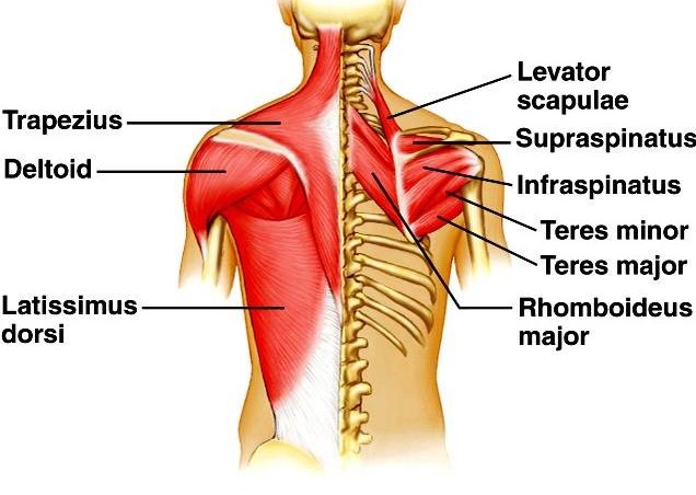
Muscle Anatomy Quiz from www.registerednursern.com And reach, pull and extend your arms and torso. For more anatomy content please follow us and visit our website: The muscles of the arm anatomical chart does an exemplary job of examining the individual muscles that make up this area of the human body, and how they work together in processes such as motion and flexibility. Within this group of back muscles you will find the latissimus dorsi, the trapezius, levator scapulae and the rhomboids. Anatomy of neck muscles diagram. #back muscle diagrams labeled #lower back muscle diagrams labeled. The muscles of the back. Broadly considered, human muscle—like the muscles of all vertebrates—is often divided into striated muscle, smooth muscle, and cardiac muscle.
Learn vocabulary, terms, and more with flashcards, games, and other study tools.
The back is the body region between the neck and the gluteal regions. Reproductive · may 27, 2021. Related posts of back muscles chart muscle anatomy review. Five pairs of lumbar spinal nerves labeled l1 to l5 branch off your spinal cord and exit through small holes between the vertebrae. #back muscle diagrams labeled #lower back muscle diagrams labeled. 12 photos of the muscles labeled front and back. This is an online quiz called back muscle diagram. A back muscle that pulls the arm down and back. Your back consists of three distinct layers of muscles, namely the superficial layer, the intermediate layer, and the deep layer. Start studying 08 back muscles label. Shoulder muscles diagram labeled : There are around 650 skeletal muscles within the typical human body. It permits movement of the body, maintains posture and circulates blood throughout the body.
This is an online quiz called back muscle diagram. Human anatomy diagrams show internal organs. They start at the top of the neck and go down to the tailbone. There are around 650 skeletal muscles within the typical human body. Another common cause of lower back and hip pain is disc injury.
Muscles Of The Pectoral Girdle And Upper Limbs Anatomy And Physiology I from s3-us-west-2.amazonaws.com And reach, pull and extend your arms and torso. Muscles make up a large part of the anatomy (structure) of the back. Anatomy of neck muscles diagram. Diagram back muscles human muscle diagram labeled the back side | humananatomybody. 12 photos of the muscles labeled front and back. The muscles of the arm anatomical chart does an exemplary job of examining the individual muscles that make up this area of the human body, and how they work together in processes such as motion and flexibility. C rnrceps brachn l unssimus dorsi k. Leg muscle anatomical structure, labeled front, side, and back view diagrams.
To learn more about the anatomy of the spine, watch this video.
It also covers some common conditions and injuries that can affect the back. Women back body parts image real. This is an online quiz called back muscle diagram. Lower back muscle diagram anatomy does degenerative disc disease affect the lower back muscle? This labeled human muscular system chart illustrates the major muscle groups in the back (posterior) view and the front (anterior) view. Anterior rami of upper thoracic the deep or intrinsic muscles of the back extend from the pelvis to the skull and are innervated by segmental. Broadly considered, human muscle—like the muscles of all vertebrates—is often divided into striated muscle, smooth muscle, and cardiac muscle. Start studying 08 back muscles label. The muscles, bones, ligaments, and tendons in the back can all be injured and cause back pain. For more anatomy content please follow us and visit our website: Another common cause of lower back and hip pain is disc injury. Back pain is one of the most common kinds of pain for adults, and muscle strains are the most common type of back pain. See more ideas about back muscles, muscle anatomy, shoulder muscle anatomy.
This article looks at the anatomy of the back, including bones, muscles, and nerves. Extrinsic and intrinsic.the back functions are many, such as to house and protect the spinal cord, hold the body and head upright, and adjust the movements of the upper and lower limbs. Start studying 08 back muscles label. Reproductive · may 27, 2021. Anterior rami of upper thoracic the deep or intrinsic muscles of the back extend from the pelvis to the skull and are innervated by segmental.
Back Muscles Attachments Nerve Supply Action Anatomy Info from anatomyinfo.com This large muscle in the back. Back muscle diagrams labeled 12 photos of the back muscle diagrams labeled back muscle diagrams labeled, lower back muscle diagrams labeled, human muscles, back muscle diagrams labeled, lower back muscle diagrams labeled. Muscle anatomy types of movement all muscles exert their force by pulling between at least two points of attachment. Learn vocabulary, terms, and more with flashcards, games, and other study tools. See more ideas about back muscles, muscle anatomy, shoulder muscle anatomy. The muscles of the lower back help stabilize, rotate, flex, and extend the spinal column, which is a bony tower of 24 vertebrae that gives the body structure and houses the spinal cord. They also attach your shoulders and pelvis to the trunk, creating a bridge between. Within this group of back muscles you will find the latissimus dorsi, the trapezius, levator scapulae and the rhomboids.
This page is about back muscles diagram.
To learn more about the anatomy of the spine, watch this video. Anterior rami of upper thoracic the deep or intrinsic muscles of the back extend from the pelvis to the skull and are innervated by segmental. We think this is the most useful anatomy picture that you need. The muscles of the back. Start studying 08 back muscles label. Other muscles that aid in shoulder movement include: 12 photos of the muscles labeled front and back. Muscles make up a large part of the anatomy (structure) of the back. Anatomynote.com found anatomy of back muscles diagram from plenty of anatomical pictures on the internet. This article looks at the anatomy of the back, including bones, muscles, and nerves. Human anatomy diagrams show internal organs. Upper back anatomy muscles anatomy drawing diagram. Studying these is an ideal first step before view the muscles of the upper and lower extremity in the diagrams below.
See more ideas about back muscles, muscle anatomy, shoulder muscle anatomy back muscle diagram. We hope this picture anatomy of back muscles diagram can help you study and research.

